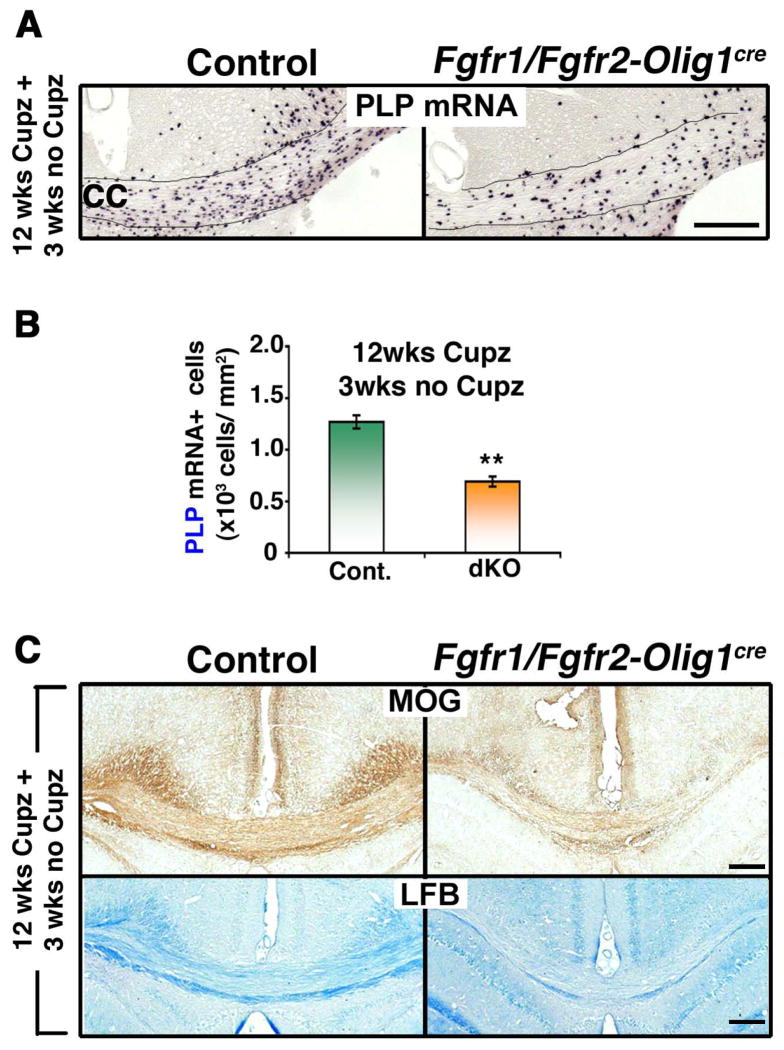
Figure 6. Repopulation of chronically demyelinated lesions by oligodendrocytes is impaired in Fgfr1/Fgfr2-Olig1Cre conditional knockout mice.
Control and Fgfr1/Fgfr2- Olig1Cre dKO mice were fed 0.25% cuprizone (Cupz) for 12 weeks (wks), followed by normal chow for 3 wks. A. Coronal sections of the brains, analyzed by PLP mRNA in situ hybridization to identify oligodendrocytes, show that after 3 wks of normal chow feeding, fewer oligodendrocytes appear in the Fgfr1/Fgfr2-Olig1Cre dKO mice compared to controls. B. Quantification of the numbers of PLP mRNA+ oligodendrocytes in the corpus callosum confirms the significant reduction in the dKOs compared to controls. Three sections per animal were counted from 3–5 mice per group. C. Coronal sections of the brain, analyzed by MOG immunolabeling and LFB staining, show reduced signals in the dKO, indicative of impaired recovery of myelin. Scale bars, 200 μm. Error bars represent S.E.M.; N=3. **p<0.01.

