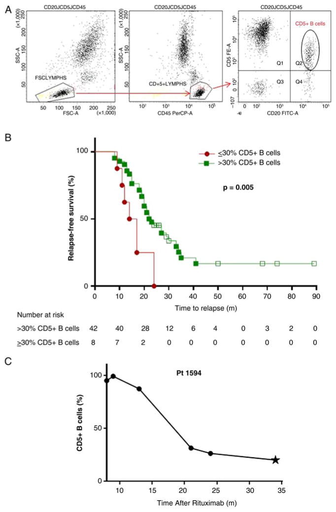
Figure 1.
Repopulation with <30% CD5+ B cells portends a shorter time to relapse than repopulation with normal levels of CD5+ B cells. (A) Gating scheme for re-analysis of clinical flow cytometry data. Whole blood was stained for a CD20 workup with the following fluorescently labelled antihuman antibodies: CD20-FITC (clone L27), CD45-PerCP (clone 2D1) and CD5-PE (clone L17F12, all from BD Biosciences, San Jose, California, USA). Cells were analysed using a FACSCanto II flow cytometer. Lymphocytes were first selected based on forward versus side scatter; CD45 was then used as a pan lymphocyte marker in combination with side scatter to identify lymphocytes in a strategy known as heterogeneous gating that is required in clinical flow cytometry core facilities.10 CD5+ B cells were defined as cells positive for both CD20 and CD5 as denoted by the oval gate. In samples used for this study, CD20 correlated well with CD19 expression (r2=0.98). (B) Relapse-free survival from the first dose of rituximab is depicted. Patients who repopulated with ≤30%CD5+ B cells (
 ) relapsed sooner than patients who repopulated with >30% CD5+ B cells (
 ; p=0.005). Open squares denote the months of follow-up for patients who did not relapse during the time of our study (n=11), with a minimum 24 months of relapse-free follow-up required. Adjusting for differences in upper respiratory involvement, the low CD5 group at B cell repopulation remained significantly associated with a shorter time to relapse from time of rituximab (p=0.002) and from time of B cell repopulation (p=0.001). (C) CD20+CD5+ B cells decrease prior to relapse. Shown is an example of a patient who repopulates with high CD5+ B cells after rituximab therapy but exhibits decreasing and then low CD5+ B cells prior to disease relapse (denoted by star).

