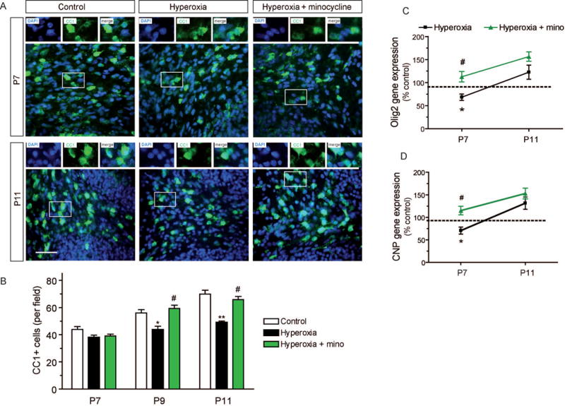
Figure 4. Perturbed maturation of oligodendroglial cells in the cerebellum after hyperoxia.

(A) Immunohistochemistry of 10 μm cerebellar sections to label mature CC1+ oligodendrocytes at P7 immediately after exposure to hyperoxia followed by recovery in room air until P9 and P11. (B) Cell numbers of mature CC1+ oligodendrocytes in the cerebellar white matter were not reduced by hyperoxia at P7. At P9 and at P11, however, CC1+ cells were significantly reduced in rats after hyperoxia. Minocycline abolished the delay of oligodendroglial maturation. (C) Gene expression of the oligodendroglial transcription factor Olig2 was significantly decreased after hyperoxia at P7, whereas minocycline improved Olig2 expression. At P11, expression levels of Olig2 were increased in minocycline treated and untreated hyperoxia rats (n.s.). (D) Gene expression levels of oligodendroglial maturation marker CNP were significantly decreased after exposure to hyperoxia at P7 as measured by qPCR. Minocycline treatment abolished the reduction of CNP expression. At P11, CNP was increased both in minocycline treated and untreated hyperoxia rats (n.s.). Scale bar = 50μm (*P<0.05 and **P<0.01 versus control, ##P<0.01 and #P<0.05 versus hyperoxia; ANOVA with consecutive t-test, (A, B) n=4, (scale bar 50 μm); (C, D) n=6).
