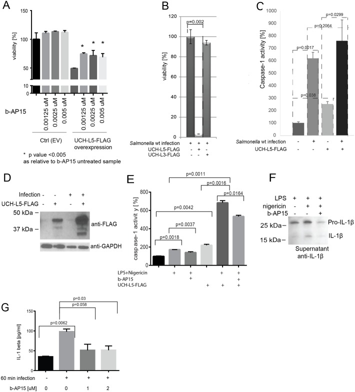
Fig 3. UCH-L5 regulates inflammasome activity in chicken macrophages.
(A-B). UCH-L5 overexpression leads to reduced cell viability in infected as well as uninfected HD11 macrophages, which depends on its catalytic activity. UCH-L5 was overexpressed in HD11 macrophages and empty vector was used as a control (Ctrl). 24-hours past overexpression, the cells were treated with b-AP15 (UCH-L5 inhibitor) used at indicated concentrations for 18 hours. The cell viability was measured by Presto Blue assay (A). Alternatively, UCH-L5 was overexpressed in HD11 macrophages for 2 days prior to infection with Salmonella Typhimurium for 1 hour. The cell viability was measured by Presto Blue assay. The p-values were calculated by using Student T test. (C) UCH-L5 overexpression leads to an increase in caspase-1 activity in HD11 macrophages. UCH-L5 was overexpressed in HD11 macrophages (UCH-L5) and empty vector was used as a control (ctrl). 24-hours past overexpression, the cells were subjected to Salmonella infection at MOI of 50:1 for 18-hours (inf) or left uninfected. The caspase-1 activity was measured by using specific inhibitor. The p-values were calculated by using Student T test. (D). Overexpressed UCH-L5 is increased in infected cells. The protein cell lysates from (C) were analyzed by SDS-PAGE, followed by anti-FLAG western blotting to demonstrate UCH-L5 expression; anti-GAPDH western blotting was used as a loading control. (E). Caspase-1 activity in HD11 macrophages is dampened upon b-AP15 inhibitor treatment. UCH-L5 was overexpressed in HD11 macrophages (UCH-L5-FLAG) and empty vector was used as a control. 24-hours past overexpression, cells were primed with 1μg/ml LPS for 4 hrs followed by treatment with 1uM b-AP15 or vehicle control (DMSO) for 15 minutes to inhibit the activity of UCH-L5. Cells were then treated with 10μM nigericin for 1 hour to induce inflammasome. The cell pellets were collected and caspase-1 activity was measured by using Z-YVAD-AFC substrate. The p-values were calculated by using Student T test. (F). Exposure of cells to b-AP15 inhibitor leads to decrease in IL-1β secretion in HD11 macrophages upon inflammasome activation. The HD11 cells were seeded on 6-well plates (1x10⁶ cells, 3 replicates each) and primed with LPS (1μg/ml) for 4 hours followed by treatment with 1uM b-AP15 (or vehicle control, DMSO) for 15 minutes. Cells were then treated with 10uM nigericin (or not) for 1 hour to induce inflammasome. Media were collected and used for detection of chicken IL-1β by western blot. (G). Exposure of chicken HD11 macrophages to b-AP15 inhibitor leads to decrease in IL-1β secretion during Salmonella infection. HD11 cells were treated with 1μM b-AP15 or DMSO (vehicle control) for 60 min. They were then infected (or not) with Salmonella Typhimurium wild-type at MOI 50:1 for 60 minutes. Media were collected for ELISA-based quantitation of chicken IL-1β (MyBioSource, Inc., USA).

