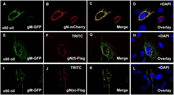
Fig 3. Transient co-expression of GPCMV gM and gN.
Tagged versions of gN and gM were transiently co-expressed in GPL cells and cellular localization patterns investigated by immunofluorescence or autofluorescence assay. Panels A-D, gMGFP and gNmCherry co-localization studies. A and B show gM and gN separately within the same cell. C is the merged image for A and B. D is the overlay of C with DAPI stain to indicate location of the nucleus. E-H, gMGFP and gN(f)FLAG co-localization with G merged image for panels E and F. H the overlay for DAPI stain with merged image G. I-L, gMGFP and gN(s)FLAG co-localization with K merged image for I and J. L overlay with DAPI staining.

