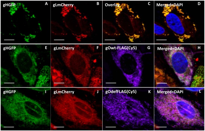
Fig 7. Transient expression and cellular co-localization of GPCMV gH, gL and gO.
Expression plasmids described in Fig 5 were used to transiently express the viral glycoproteins in GPL cells. Panels A-D, gHGFP and gLmCherry co-expression. A and B cellular location of gH and gL within the same cell respectively. C, overlay of A and B. D, merge of overlay and DAPI stain to indicate location of nucleus. Panels E-H, gHGFP, gLmCherry and gOFLAG. Individual cellular localization panels E, F and G. Merged panel H, shows co-localization of all three protein and DAPI stained nucleus. Panels I-L are for gHGFP, gLmCherry and gO(def)FLAG. Individual panels I, J and K. Merged image (L) for all three panels plus DAPI stained nucleus.

