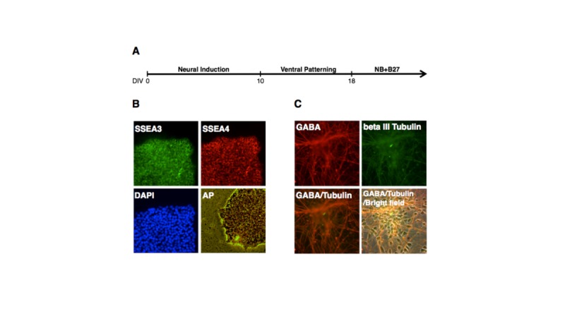
Fig 1. Neuronal differentiation of H1 hESCs.

A. Schematic diagram for differentiating H1 hESCs into cortical neurons. B. Immunostaining [using antibodies reacting with stage-specific embryonic antigen-3 (SSEA3) and antigen-4 (SSEA-4)], nuclear staining [using 4',6-diamidino-2-phenylindole (DAPI) which is a fluorescent stain that binds strongly to A-T rich regions in DNA], and alkaline phosphatase (AP) staining (using a fluorescent substrate for AP for characterizing pluripotent stem cells) of H1 colonies before neuronal differentiation. C. Immunostaining of hESCs-derived neurons at days in dish (DIV) 32 using antibodies reacting with GABA and beta III tubulin, which are the biomarkers for GABA neurons.
