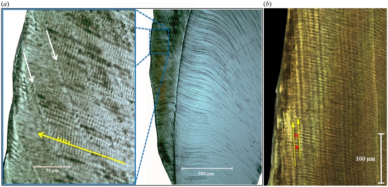
Fig 1. Appearance of tooth enamel in histological sections.
(a) An orangutan (Pongo pygmaeus pygmaeus) molar. Dentine is toward the right, enamel surface to the left. Cusp tips are toward the top. In the inset (adapted from [16]), individual enamel prisms (yellow arrow) run from the dentine to the enamel surface, with daily cross striations (yellow hash-marks) running across prism long axes. Striae of Retzius (white arrows) run obliquely from outer enamel to the enamel-dentine junction. RP (11 days here) can be determined by counting cross striations between successive striae of Retzius. (b) A diademed sifaka (Propithecus diadema) molar, whose RP = 3 days (yellow arrows = striae of Retzius, with 3 cross-striations visible between them; red arrows demarcate transition between striations).

