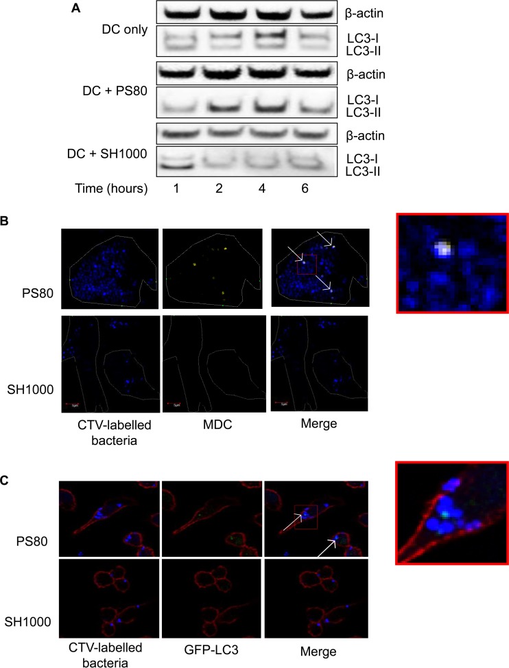
FIG 4.
S. aureus strain PS80 inhibits normal autophagic flux in phagocytes. (A) BMDCs were infected with S. aureus strain PS80 or SH1000. At the indicated time points, cells were lysed, and expression of LC3 was analyzed by Western immunoblotting. Bands show the conversion of LC3-I to LC3-II. The level of β-actin was measured as a loading control. Representative blots from 3 independent experiments are shown. (B) Six hours after infection with CTV-labeled bacteria, BMDCs were stained with MDC and fixed to be viewed under a fluorescence microscope. Blue, bacteria; yellow, MDC. White arrows indicate colocalization of bacteria and LC3-II. (C) Three hours after infection with CTV-labeled bacteria, GFP-LC3 iBMMs were fixed, permeabilized, and stained with phalloidin for viewing under a fluorescence microscope. Blue, bacteria; green, LC3; red, phalloidin. White arrows indicate colocalization of bacteria and LC3-II. See also the enlarged images, which show the extent of colocalization.

