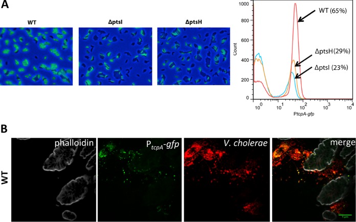
FIG 4.
A smaller fraction of cells express tcpA in PTS mutants. (A) GFP expression from a chromosomal PtcpA-gfp reporter in the wt and ptsI and ptsH deletion mutants after growth under AKI conditions was monitored with epifluorescence microscopy (left) or flow cytometry (right). The figure shows representative results from three replicates. (B) Heterogeneous expression of tcpA in wt V. cholerae is also seen during infection of suckling mice. V. cholerae cells in the small intestine (red) were detected by immunofluorescence, and tcpA expression (green) was detected from the chromosomal PtcpA-gfp reporter. Tissue sections were counterstained with phalloidin (gray) to visualize actin.

