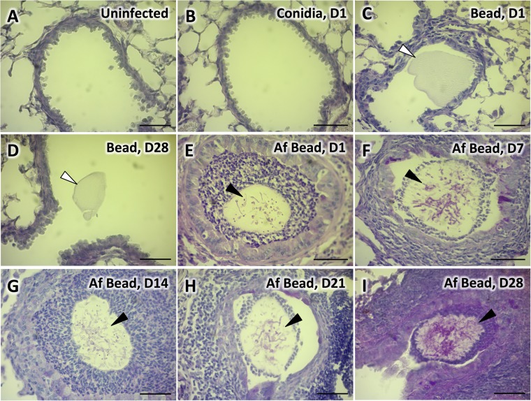
FIG 2.
Colonization of airways with A. fumigatus beads. Histopathology of PAS-stained lung tissue of uninfected C57BL/6 mice (A) or mice infected with conidia in suspension 24 h postinfection (B), sterile beads 24 h postinfection (C), sterile beads 28 days postinfection (D), A. fumigatus beads 24 h postinfection (E), A. fumigatus beads 7 days postinfection (F), A. fumigatus beads 14 days postinfection (G), A. fumigatus beads 21 days postinfection (H), or A. fumigatus beads 28 days postinfection (I); images were taken with a 40× objective lens. White arrows indicate sterile beads. Black arrows indicate PAS-positive hyphae of A. fumigatus within agar beads. Bars, 50 μm. Histopathology of PAS-stained lung tissue of C57BL/6 mice infected with conidia in suspension or sterile beads on days 7, 14, and 21 days postinfection can be found in Fig. S1 in the supplemental material.

