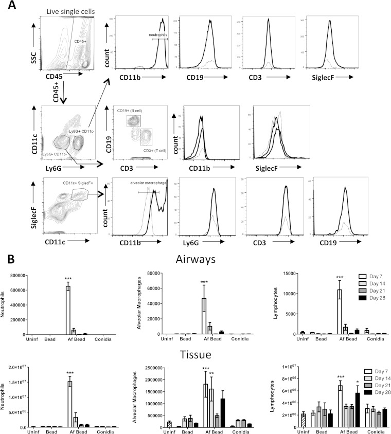
FIG 4.
(A) Gating strategy for leukocyte differential analysis by flow cytometry. Mouse lungs were digested by collagenase, and cells were stained with fluorescently labeled antibodies. Doublet cells and dead cells, determined using a fixable viability dye, were excluded. Histograms show the cell surface staining for a fully stained sample (solid line), as well as the “fluorescence minus one” control (dotted line) for the corresponding channel. (B) A. fumigatus beads induce pulmonary leukocyte recruitment. Total numbers of the indicated leukocyte populations were determined by flow cytometry analysis of BAL fluid (airways) and lung (tissue) samples. Bars represent the means ± standard errors of the means from 4 to 8 mice per group per time point. *, P < 0.05; **, P < 0.01; ***, P < 0.001, compared to uninfected mice.

