FIG 6.
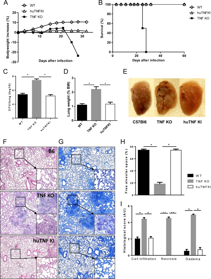
Human TNF sustains host control of acute M. tuberculosis infection in huTNF KI mice. (A and B) WT, TNF KO, and huTNF KI mice were infected with M. tuberculosis H37Rv (i.n., 1,000 CFU per lung) and their body weights (A) (n = 19 for WT mice, n = 22 for huTNF KI mice, n = 5 for TNF KO mice) and survival (B) (n = 8 to 10) were monitored. Data are mean values from one experiment and are representative of those from two independent experiments. (C and D) Pulmonary bacterial loads (numbers of CFU) (C) and lung weights (D) in WT, huTNF KI, and TNF KO animals were measured at 4 weeks postinfection (n = 5 mice per group). BW, body weight. (E) Macroscopic examination of infected lungs. (F and G) Microscopic examination of WT, huTNF KI, and TNF KO mice on day 28 after infection using H&E staining (F) or Ziehl-Neelsen staining (G). Magnifications, ×50, ×200 (insets in panel F), and ×400 (insets in panel G). (H and I) Semiquantitative scores of inflammatory cell infiltration into the lung, necrosis, and edema on day 28 (n = 5/group). Bar graphs show means ± SEMs. AU, arbitrary units. *, P < 0.05 versus WT; **, P < 0.01 versus WT.

