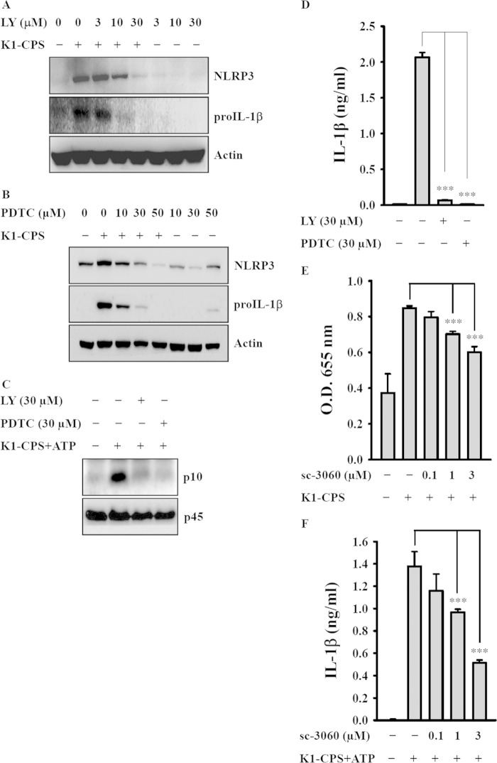
FIG 7.
Effect of phosphatidylinositol 3 (PI3)-kinase and NF-κB on K1-CPS-mediated NLRP3 inflammasome activation. (A and B) J774A.1 macrophages were incubated for 30 min with or without LY294002 (LY) (A) or PDTC (B) and then for 6 h with or without K1-CPS. The levels of NLRP3 and pro-IL-1β in the cells were measured by Western blotting. (C and D) J774A.1 macrophages were incubated for 30 min with or without LY or PDTC, for 5.5 h with or without 1 μg/ml of K1-CPS, and then for 30 min with or without 5 mM ATP. The levels of activated caspase-1 (p10) in the cells (C) and IL-1β in the culture medium (D) were measured by Western blotting and ELISA, respectively. (E) J-Blue cells were incubated for 30 min with or without sc-3060 and then for 24 h with or without 1 μg/ml of K1-CPS. The activation levels of NF-κB were measured by an NF-κB reporter assay. (F) J774A.1 macrophages were incubated for 30 min with or without sc-3060, for 5.5 h with or without 1 μg/ml of K1-CPS, and then for 30 min with or without 5 mM ATP. The levels of IL-1β in the culture medium were measured by ELISA. In panels A to C, the results are representative of three separate experiments. In panels D to F, the data are expressed as the means ± SD from three separate experiments. ***, P < 0.001.

