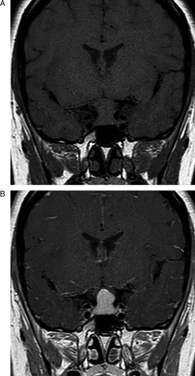
Figure 1.

Initial pituitary MRI revealing pituitary gland enlargement. (A) unenhanced coronal image showing an enlarged pituitary gland with supra- and perisellar expansion and compression of the optic chiasm. (B) Homogeneous enhancement of the pituitary lesion after Gd-DTPA.

 This work is licensed under a