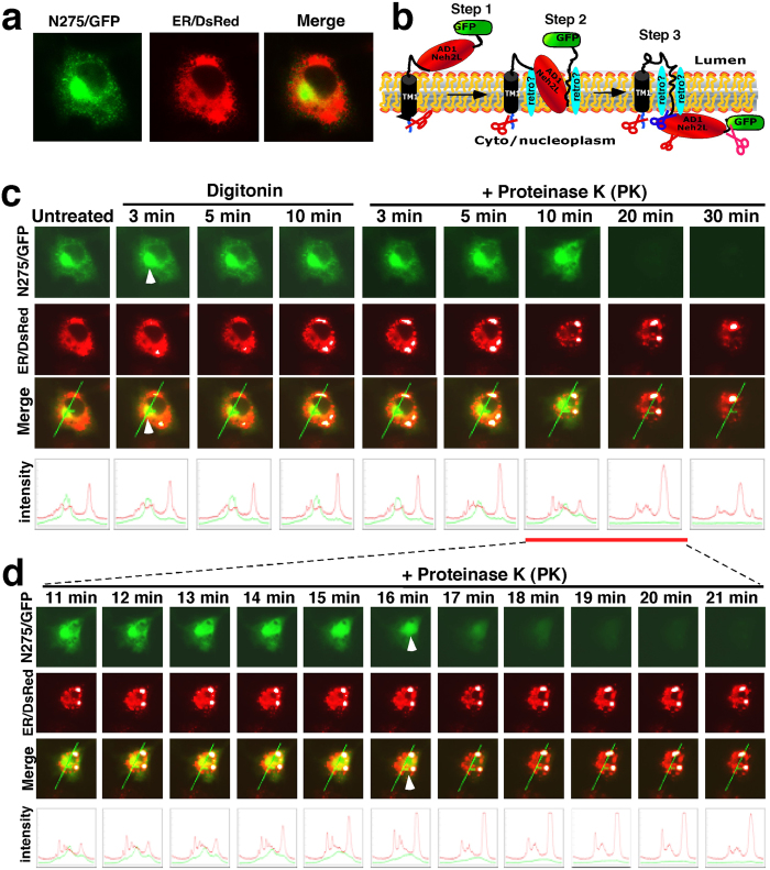
Figure 2. Live-cell imaging of N275/GFP that moves dynamically out of the ER membrane into the cytoplasm.
(a) Localization of N275/GFP (in which the N275 portion contains the entire NTD and most of AD1 from Nrf1) within and around the ER. The merged images of Nrf1/GFP with ER/DsRed are also shown. (b) The putative dynamic membrane-topologies of N275/GFP are shown schematically. Within N275/GFP, the AD1 of Nrf1 has been fused C-terminally by GFP, which is postulated to face the ER luminal side of membranes during the initial topogenic vectorial process (see refs 25,26). (c) COS-1 cells co-expressing N275/GFP and the ER/DsRed marker were subjected to live-cell imaging combined with the in vivo membrane protease protection assay. During co-incubation of the cells with digitonon (20 μg/ml) and proteinase K (PK, 50 μg/ml) for 30 min, real-time live-cell images were acquired using the Leica DMI-6000 microscopy system. The merged images of Nrf1/GFP with ER/DsRed are placed (on the third raw of panels), whereas changes in the intensity of their signals are shown graphically (bottom). Overall, the images shown herein are a representative of at least three independent experiments undertaken on separate occasions that were each performed in triplicate (n = 9). (d) Additional live-cell imaging of Nrf1/GFP was acquired from 11 to 21 min after co-incubation of PK with digitonin, as described above.

