
Fig. 1.
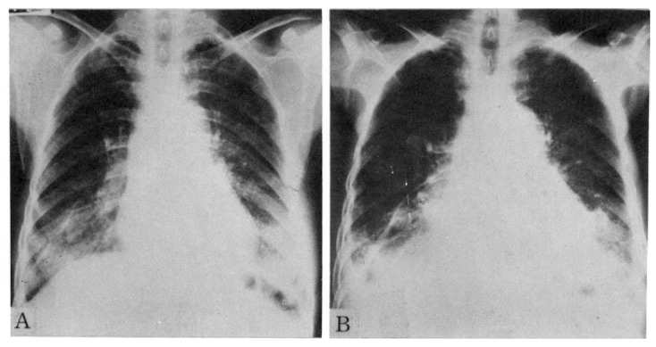
Serial chest radiographs of 60-year-old female patient.
A. The Initial radiograph shows bilateral diffuse ground glass appearing density, especially in the lower lung fields.
B. Radiograph obtained one year later A shows both aggravated lower lung field lesions. Note bilateral pleural effusion and increased heart size as compared with radiograph A.