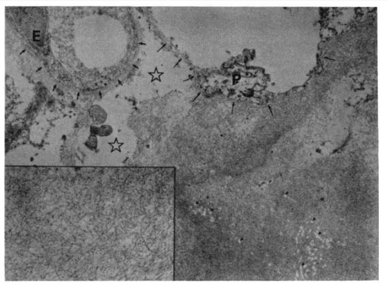
Fig. 6.

Electron microscopic observation of the lung reveals massive deposition of fibrillar material in the alveolar septum. The alveolar basement membrane (long arrows) is incorporated into the amyloid substance. Retractile white spots and fibers (tiny arrowheads) in the amyloid pool are preexisting collagen fibers. Free space (stars) in the interstitium is due to interstitial edema. Alveolar epithelial cells (P) are artifactually deformed. (E: endothelial cells; smaller arrows: capillary basement membrane). (Bar represents 1 um, ×14,000)
(Inset) Magnification of the amyloid substance reveals nonbranching fibrills of approximately 100 nm in length and 10 nm in diameter. Granular deposits, less than 10 nm in diameter, are scattered also. (Bar represents 0.1 um, ×56,000).
