
Fig. 2.
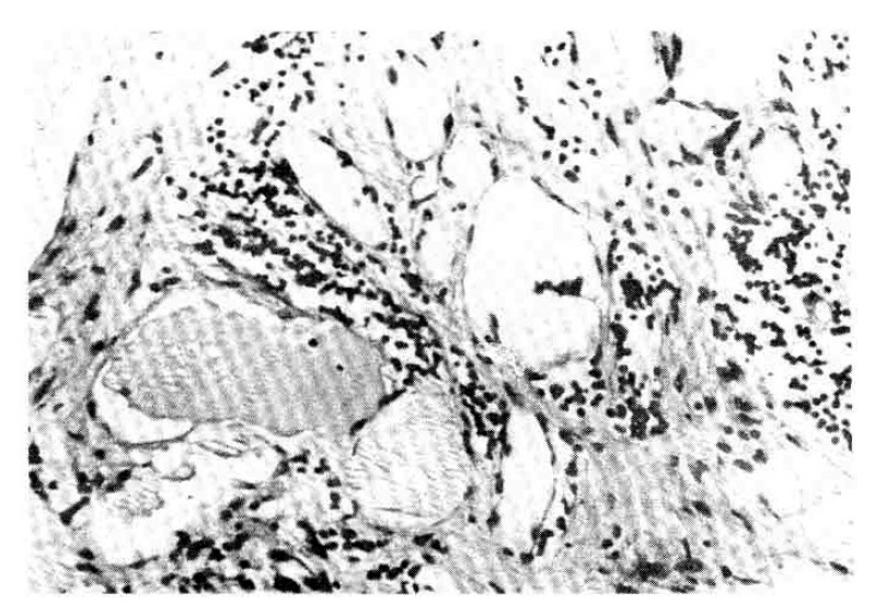
The cysts are lined by flattened endothelial cells and contain pale eosinophilic fluid with lymphocytes in the lumens. Scattered infiltrates of lymphocytes in the stroma are present (H-E stain, × 400).
Official websites use .gov
A
.gov website belongs to an official
government organization in the United States.
Secure .gov websites use HTTPS
A lock (
) or https:// means you've safely
connected to the .gov website. Share sensitive
information only on official, secure websites.
The cysts are lined by flattened endothelial cells and contain pale eosinophilic fluid with lymphocytes in the lumens. Scattered infiltrates of lymphocytes in the stroma are present (H-E stain, × 400).