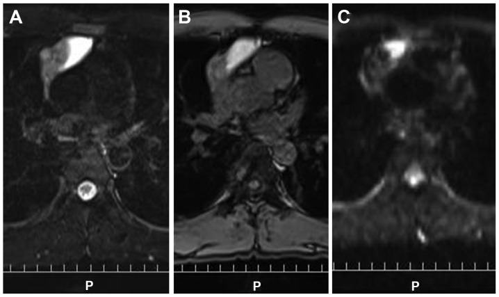
Figure 4.
The left side of the tumor shows a high signal on (A) T2-weighted imaging and (B) fat suppression T1-weighted imaging, suggesting high protein and serum content of the cystic structure. The remaining portion of the tumor shows a mild high signal on (C) diffusion weighted images and T2-weighted images, suggesting a solid component.

