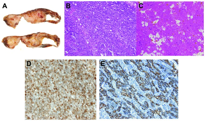
Figure 5.
(A) Macroscopically, the resected specimen was composed of a solid mass with massive necrosis and a cystic space; (B) the mass in the right lobe is composed of epithelial cells admixed with lymphocytes; (C) the tumor included prominent degenerative and necrotic areas; (D) on CD5 immunohistochemical staining, the presence of T lymphocytes was demonstrated; (E) immunohistochemical staining was strongly positive for cytokeratin (AE1/AE3).

