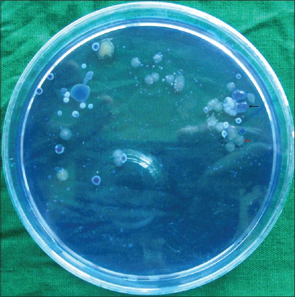
Figure 2.

Growth of Streptococcus mutans (shown in black arrow) seen as raised, convex, opaque, pale-blue colonies that are granular (i.e., “frosted glass”) in appearance, exhibiting a glistening bubble on the surface due to excessive synthesis of glucan from sucrose and Streptococcus salivaris (shown in red arrow) seen as large, pale-blue, mucoid colonies that are glistening (i.e. “gum-drop”) in appearance on Mitis Salivarius agar (original)
