
Figure 1.
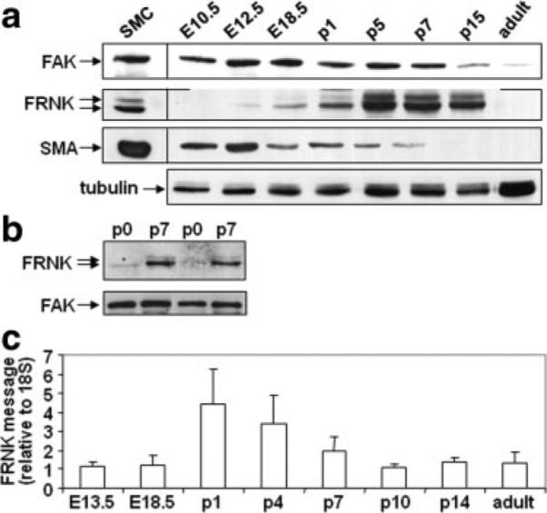
Endogenous cardiac FRNK expression during heart development. a and b, Western blots of cardiac lysate from indicated embryonic (E), postnatal day (p), or adult C57black6 mice (a) or cardiomyocytes isolated from P0 or P7 neonatal rat hearts (b). Western blots are representative of at least 3 separate experiments. c, Quantitative RT-PCR for FRNK message in C57black6 hearts. FRNK levels were normalized to 18S and presented as fold change over message levels detected at E13.5±SEM (n=at least 4 for each time point).