
Figure 3.
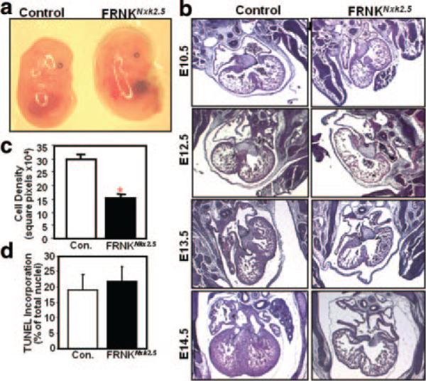
Histological and morphological analysis of FRNKNkx2.5 mice. a, Gross morphology of genetic control and FRNKNkx2.5 embryos at E14.5. b, Hematoxylin/eosin-stained sections of hearts from the indicated embryonic day. c, Cellular density was quantified in serial sections from E14.5 genetic control and FRNKNkx2.5 hearts. d, Ventricle sections from E13.5 genetic control and FRNKNkx2.5 mice were costained with FragEL and cardiac troponin T to identify apoptotic cardiomyocytes. Graphical data are expressed as means±SEM (n=6 for each condition; *P<0.01).