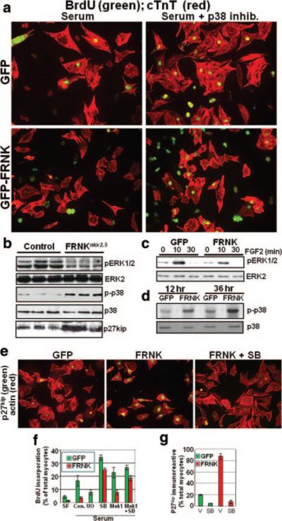
Figure 5.
FRNK expression attenuates DNA synthesis and promotes cell cycle withdrawal in a p38-dependent fashion. a, BrdUrd incorporation in isolated cardiomyocytes (P0) infected with GFP- or GFP-FRNK adenovirus (10 multiplicities of infection). Cells were maintained in serum-containing medium and treated with vehicle or SB203580 (10 μmol/L) as indicated. Costaining with anti–cardiac troponin T (cTnT) was performed to identify cardiomyocytes. b, Western blot analysis in cardiac lysates from E13.5 FRNKloxP and FRNKNkx2.5 embryos. c and d, Rat neonatal cardiomyocytes (P0) plated on fibronectin were infected with GFP- or GFP-FRNK adenovirus (10 multiplicities of infection), replaced with serum-free media, and treated with FGF-2 (c) or maintained in serum-containing conditions (d) for the indicated times before immunoblotting. See Online Figure VI (a through c) for densitometric quantification of 3 separate experiments. e, Isolated cardiomyocytes, treated as described for a, were processed by immunocytochemistry for p27kip (myocytes are identified by striated actin visualized by phalloidin). f and g, Quantification of BrdUrd- and p27kip-positive cardiomyocytes (means±SEM; n=3; minimum of 300 cells/condition). Additional representative images are provided in Online Figure VIII.

