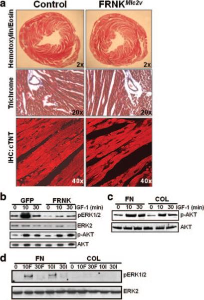
Figure 6.
Anabolic cardiac growth and AKT signaling are not affected by FRNK expression. a, Low- and high-power images of cross-sections of 8-week-old FRNKloxp and FRNKMlc2v hearts stained with trichrome (blue corresponds to collagen) (top and middle) or with an anti–cardiac troponin T antibody (bottom). b and c, Rat neonatal cardiomyocytes were infected as described above or plated on fibronectin (FN) or collagen (COL) (10 μg/mL) and treated with IGF-1 or FGF-2 for the indicated times. Lysates were immunoblotted for phospho- and total ERK and AKT. Western data are representative of at least 3 separate experiments. See Online Figure VI for quantification.

