
Fig. 4.
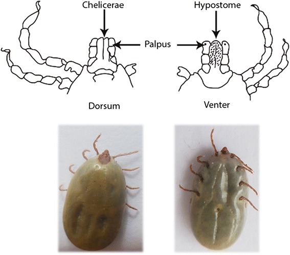
Photo of a tick and schematic representation of the capitulum. Dorsal and ventral morphology of the mouthpart of Ixodidae family ticks. On the dorsum it is possible to observe the chelicerae while the venter displays the hypostome. The palpus is observable on both sides (dorsum and venter). The hypostome is responsible for the dermal and epidermal damage (rupture of local blood vessels) during the tick’s feeding