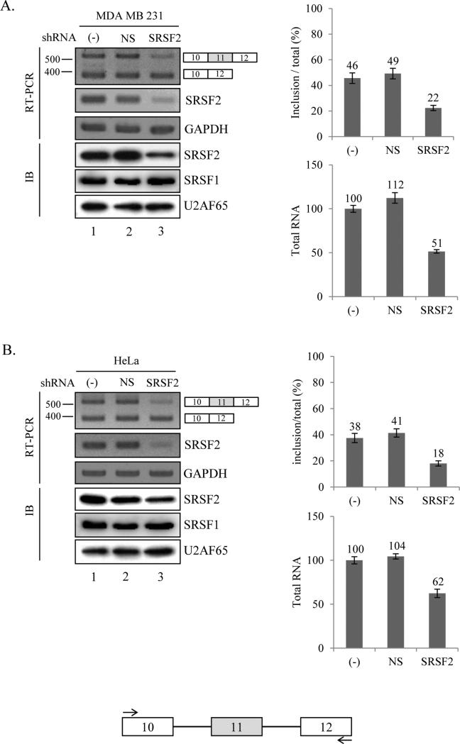
Figure 1.
Knockdown of SRSF2 with shRNA reduced exon 11 inclusion of Ron pre-mRNA. RT-PCR analysis of endogeneous Ron exon 11 alternative splicing with RNAs extracted from non-treated cells, non-silencing shRNA virus treated cells and SRSF2 shRNA virus treated cells are shown. Primers are located at exon 10 and exon 12 as shown with arrows. MDA MB 231 (A) and HeLa (B) cell lines are tested here. GAPDH is used as a control. Quantitative results are shown at right panels. Immunoblotting analysis in these cells using anti-SRSF2, anti-SRSF1 and anti-U2AF65 antibody are shown.

