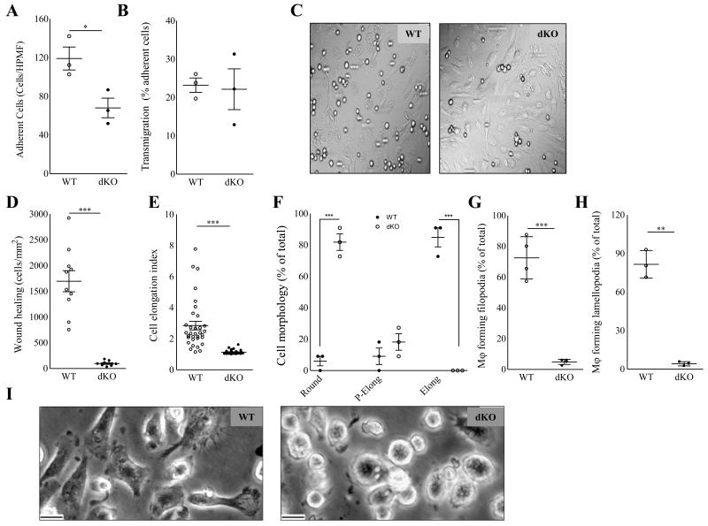
Figure 4.
Hck/Fgr dKO macrophages display impaired two dimensional directional migration and aberrant morphology. Adhesion (A) but not transmigration (B) across monolayers of inflamed endothelium in vitro is reduced in dKO BMDM (n=3, 6 High Power Microscopic Field (HPF) quantifications per sample). (C). Representative differential Interference Contrast microscopy HPMF pictures of BMDM adherent to inflamed endothelium in vitro. D. dKO macrophages display 94% reduced two dimensional migration and wound healing capacity in vitro (n=3, 5 area quantifications per replicate). E+F. Morphology of peritoneal macrophages (PEM) cultured for 36h (n=5, 100 cells per replicate). E. Mean elongation index (EI, defined as the ratio of cell length to cell breadth). F. Percentage of rounded (EI<1.2), partially elongated cell (1.2<EI<,2) and elongated (EI>2) and as judged by the elongation index (EI). G+H. Percentage of PEM cultured for 16 hours, forming filopodia (G) and lamellipodia (H) (n=5, 100 cells per replicate). I. Representative pictures depicting rounded cell morphology in dKO compared to WT PEM. (Scale bar: 10μm). WT: open circles; dKO: filled circles; *P≤0.05, ***P≤0.001.

