Figure 5.
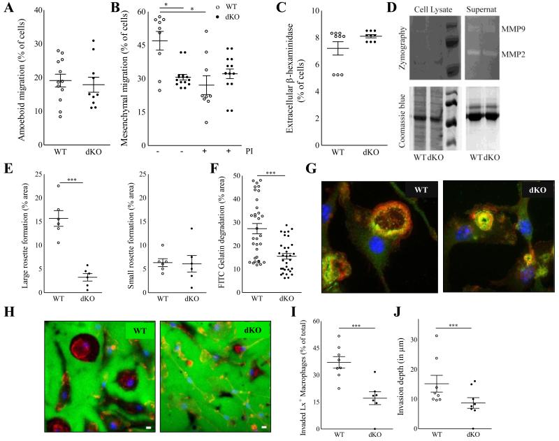
Hck/Fgr deficient macrophages have impaired three-dimensional migration capacity in vitro and in vivo. Hck/Fgr-deficient BMDM displayed normal amoeboid (mean ± S.E.M. of n=12) (A) but reduced mesenchymal migration across Matrigel transwells where proteinase inhibitors (PI) inhibited WT but not dKO BMDM migration (mean ± S.E.M.; n=9-13) (B). β-hexosaminidase was released at similar levels in WT and dKO BMDM (mean ± S.D. of n=3, in triplicate) (C). Metalloproteinases MMP-2 and MMP-9 secretion, assessed by gelatin zymograph, is unaffected in dKO BMDM (D). DKO BMDM form less and smaller podosome rosettes than WT controls (n=5) (E). Representative pictures illustrating large podosome rosettes (left panel) in WT BMDM and small podosome rosettes (right panel) in dKO BMDM, 100X magnification. (F). FITC-gelatin degradation is reduced in dKO BMDM (n=3). (G+H). Representative pictures of BMDM showing fewer and smaller gelatin proteolysis areas (in dark) colocalising with podosome rosettes in dKO BMDM compared to WT controls. (Blue for cell nuclei, red for F-actin in F and H. Green for Vinculin in F, and FITC-gelatin in H). Likewise, re-inspection of the in vivo latex bead aided monocyte/macrophage tracking experiment showed that 24h after labeling the portion of Latex bead+ plaque contained macrophages that had invaded into the atheroma (defined as located at > 3 μm from the endothelium) was significantly reduced in dKO chimeras (n=8) (I), while also the average plaque invasion depth of latex+ labeled macrophages was seen to be reduced in these mice (J). WT: open circles; dKO: filled circles; *P≤0.05, **P≤0.01, ***P≤0.001.

