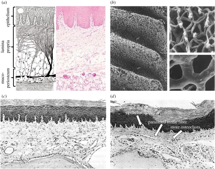
Figure 1.
(a) Schematic diagram (left) and histological diagram of the healthy mucosal anatomy [34]; (b) SEM images of the vascular network within the rabbit palatine mucosa by corrosion casts [35]; (c) histological image of the mouse mucosa underneath the denture without occlusal load [13] and (d) histological image of the mouse mucosa beneath a denture [13].

