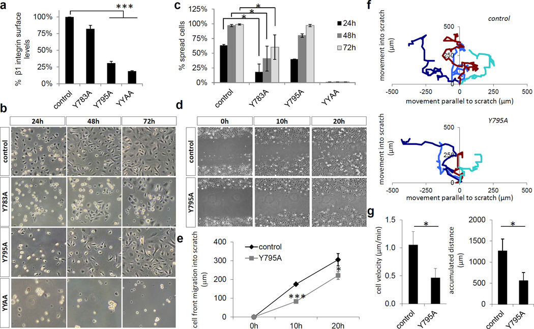
Figure 4. Adhesion and migration of β1 Y-to-A keratinocytes.
(a) Epidermis-derived single cell suspensions of normal and β1 Y-to-A epidermis were assayed for β1 integrin surface expression (mean ± SD; n=4; ***P<0.0001 vs. control). (b) Adhesion of normal and β1 Y-to-A keratinocytes to fibronectin and collagen coated tissue culture plastic over the indicated time period. (c) Spreading of normal and β1 Y-to-A keratinocytes on fibronectin and collagen coated tissue culture plastic (mean ± SD; n=3; *P<0.05 vs. control). (d) Scratch wounding of a confluent monolayer of normal and β1 Y795A keratinocytes. (e) Quantification of cell front migration into a scratch wound (mean ± SD; n=3; *P<0.05, ***P<0.0001 vs. control). (f) Assessment of migratory directionality. (g) Quantification of keratinocyte migratory velocity and accumulated distance (mean ± SD; n=3; *P<0.05 vs. control).

