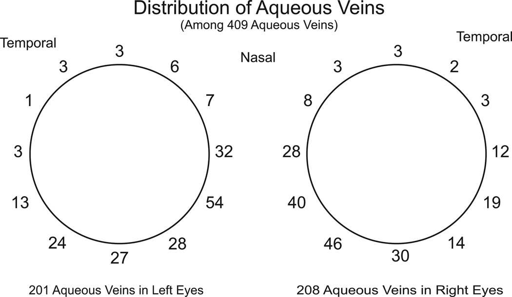
Fig. 3.
Distribution of visible aqueous veins. Two to three aqueous veins are typically visible in an eye although there may be a maximum of four to six. Distribution is highly assymetric with the majority of visible aqueous veins at or below the horizontal midline, the greatest number being present in the nasal quadrant. Derived from data of De Vries (1947).

