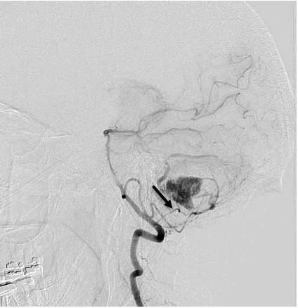
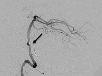
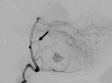
Figure 1. Angiographic images of the hemangioblastoma and arterial feeders into the vascular tumor bed. A. Pre-embolization lateral view and feeding arteries from posterior inferior cerebellar artery (arrow indicating the feeder of interest). B. The entrapped microcatheter during retraction in lateral view (arrow indicates straightening of posterior inferior cerebellar artery loop and lack of arterial flow). C. The entrapped microcatheter in relaxed position in lateral view (arrow indicates reconstitution of posterior inferior cerebellar artery loop and resteroration of arterial flow).


