
Fig. 6.
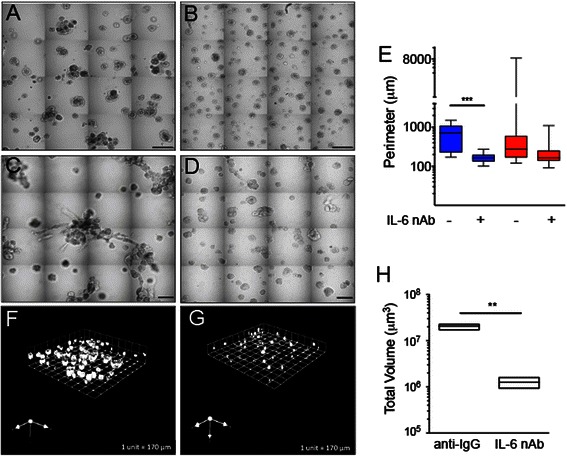
IL-6 nAb inhibits CAF-stimulated MCF10.DCIS structure growth and results in altered morphology. Contiguous 16-tile DIC image of CAFs: WS12Ti (A, B) or CAF40TKi (c, d) co-cultured with MCF10.DCIS cells for 8 days in the presence of isotype matched anti-IgG (a, c) or 1 μg /ml IL-6 nAb (b, d). e Perimeter measurements from DCIS structures in MAME culture in the presence of anti-IgG or IL-6 nAb (N = 100-140 measurements from 6 contiguous tiled DIC images (N = 3). Fluorescent tiled images of CAF40TKi fibroblasts (unlabeled) cultured with MCF10.DCIS-RFP cells in the presence of anti-IgG (f) or 1 μg /ml IL-6 nAb (g). h Quantification of total volume of DCIS-RFP structures in co-culture with CAF40TKi fibroblasts (ratio-paired t-test, N = 3). ***P ≤ 0.001, **P ≤ 0.01; Student’s t-test; mean ± SD