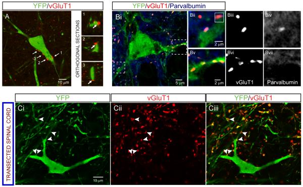
Figure 3. Anatomical evidence that primary afferents project to dI3 INs.
(A) Left: YFP+ dI3 IN with vGluT1+ boutons apposed (labeled by arrows). Right: Orthogonal sections confirming apposition of boutons labeled 1-3.
(B) vGluT1+ boutons that are PV+ and PVnull on a YFP+ dI3 IN from a P7 spinal cord. Boutons in dashed boxes are magnified in Bii-Bvii. Inset in 3Bii depicts orthogonal sections of the PV+/YFP+ bouton in the Y-Z plane.
(C) vGluT1+ boutons (arrowheads) on YFP+ dI3 IN from chronically transected spinal cord confirm that they do not originate from supraspinal descending inputs.
All images from Isl1-YFP mice.

