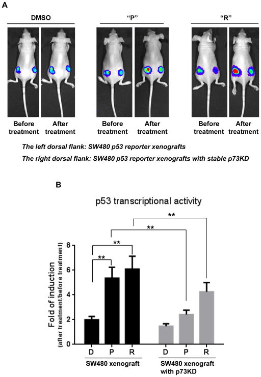
Figure 6. Prodigiosin and compound R induce p53-dependent transcriptional activity in vivo in a mutant p53-expressing human colon tumor xenograft.
(A), Tumor cells (SW480 p53 reporter cells and SW480 p53 reporter cells with stable p73KD) were inoculated into nude mice. After 1 week, mice were treated with 5mg/kg of “P”, “R” or DMSO. Before treatment, bioluminescence imaging was performed using the IVIS imaging system. After 12 hr of drug or control treatment, imaging was performed again. (B), The bar graph indicates the quantitative data for luciferase signal from 6 mice in each group. The graph shows the fold of induction of p53 transcriptional activity (After treatment/before treatment) in the “P”, “R” or DMSO groups. The mean fold of induction ± SEM is shown. (** P < 0.01, “P” vs “D” or “R” vs “D” by unpaired t-test; ** P < 0.01, SW480 vs SW480 p73KD in “P” or “R” treatment by paired t-test).

