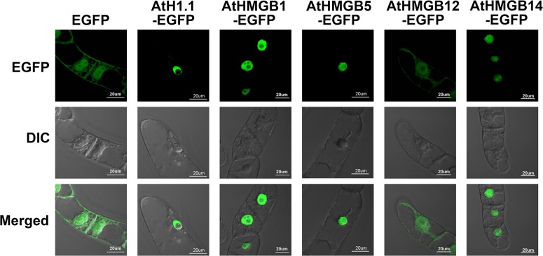
Fig 1. Interphase localization of EGFP tagged AtHMGB and AtH1.1 proteins.
Live transgenic tobacco BY-2 cells expressing free EGFP, AtH1.1-EGFP, AtHMGB1-EGFP, AtHMGB5-EGFP, AtHMGB12-EGFP, and AtHMGB14-EGFP. Confocal microscope was used to study the localization of the EGFP tagged AtHMGB proteins at interphase without fixation. Top row: EGFP signal; middle row: Differential Interference Contrast (DIC); low row: merged image.

