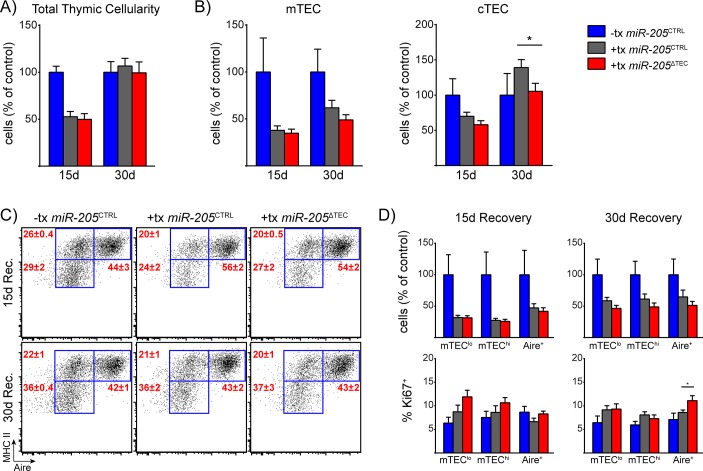
Fig 7. Radiation-induced thymic stress does not reveal a role for miR-205 in TECs.
(A) 6-week old mice were exposed to sub-lethal total body irradiation and then harvested after either 15 or 30 days of recovery to enumerate total thymic cellularity. (B) Enumeration of total mTEC and cTEC cellularity from mice shown in (A). mTECs were defined as CD45-, EpCAM+, Ly51-, MHC II+ events. cTECs were defined as CD45-, EpCAM+, Ly51+, MHC II+ events. (C) Subset composition of mTECs was assessed by flow cytometry of mTECs as defined in (B). (D) Quantification of mTEC cellularity and assessment of the proliferation marker Ki67 for the mTEC subsets shown in (C). Data in (A-D) are shown as mean ±SEM of 10 samples per group pooled from two independent experiments. Total cellularity plotted as percent of untreated miR-205 CTRL mice to allow for direct comparison between the two timepoints. Blue bars indicate untreated miR-205 CTRL mice, gray bars indicate treated miR-205 CTRL mice, and red bars indicate treated miR-205 ΔTEC mice. * denotes p≤0.05, Student’s t-test.

