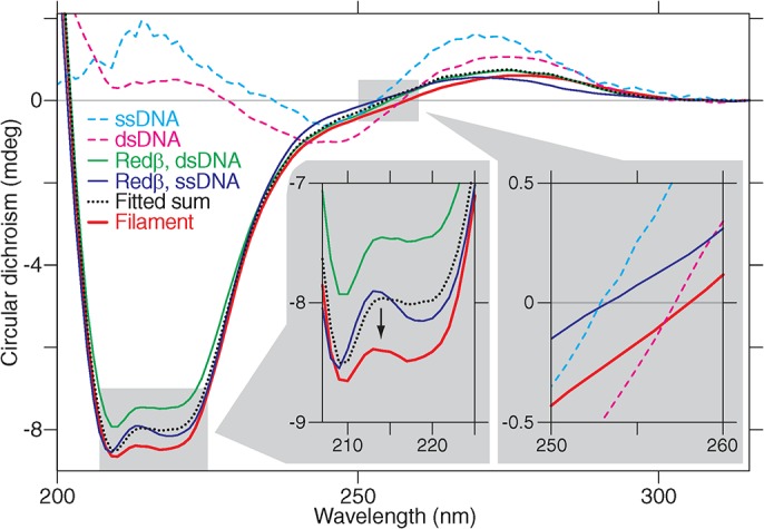
Fig 4. Circular dichroism (CD) suggests a structural change of Redβ upon filament formation.

Circular dichroism spectra show a decrease for the nucleoprotein filament (left inset shows a magnified view). The dotted black line is a fit of arbitrary contributions of the spectra of ssDNA, dsDNA, free Redβ, Redβ +ssDNA, and Redβ +dsDNA such that the concurrently recorded absorbance spectra have a maximal overlap with that of the filament (see Fig C in S1 Text for details). In this manner, the decreased circular dichroism cannot be accounted for and, therefore, must stem from a structural change. The right inset shows a bathochromic circular dichroism shift between ssDNA, dsDNA, and filament.
