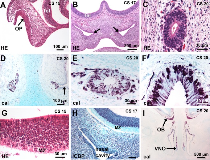
Fig 1. Development of the human olfactory system.
(A-F) Development of the OE and VNE. (A) The arrow points to the invaginated OP in front of the telencephalon. (B) The nasal cavities have developed and the presumptive VNO is first delineated (arrows). (C) A transverse section of a VNO shows a small tube lined with a pseudostratified epithelium. (D, E) The entry of the VNO (arrow) is lined with a respiratory epithelium; calretinin-positive cells are present in the VNE and in the cell-stream leaving the VNO. (F) Many calretinin-positive cells are present in the OE. (G-I) Development of the OB. (G) Higher magnification of A shows the basal telencephalon consists of a thick proliferating ventricular zone and a narrow marginal zone. (H) ICBP immunostaining (purple nuclei) shows that the MZ has developed. (I) A low magnification of a coronal section at CS 20 shows that the olfactory fibres have reached the telencephalon and form a distinct layer at the surface of the developing OB; OBs are present with a peripheral fibre layer, a differentiating layer and a ventricular layer. Abbreviations: (cal) calretinin immunostaining, (CS) Carnegie stage, (HE) hematoxylin-eosin staining, (MZ) marginal zone, (OB) olfactory bulb, (OP) olfactory placode, (Tel) telencephalon.

