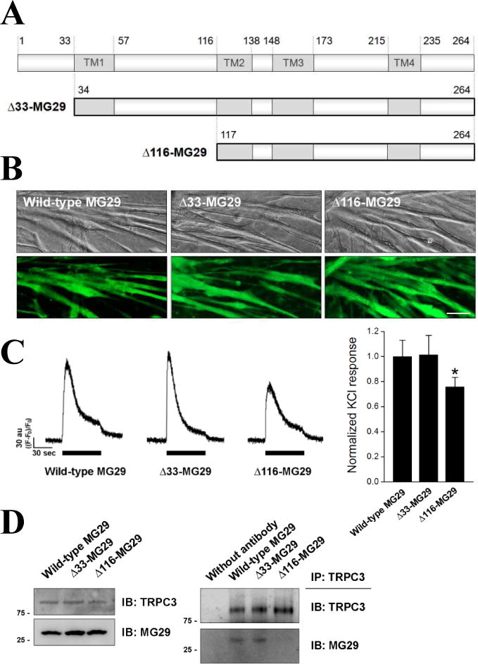
Figure 2. A reduction in Ca2+ transients in response to membrane depolarization, and the disruption of the binding between endogenous MG29 and TRPC3 in mouse primary skeletal myotubes expressing Δ116-MG29.

(A) Schematic diagram of two MG29 deletion mutants: Δ33-MG29 and Δ116-MG29. (B) Successful expressions of each mutant in mouse primary skeletal myotubes were confirmed by the presence of the GFP signal. Bar represents 50 μm. (C) KCl inducing membrane depolarization was applied to the myotubes expressing each mutant. Histograms are shown for normalized peak amplitude to the mean value of those from wild-type MG29 (92 wild-type MG29, 90 Δ33-MG29, or 95 Δ116-MG29 myotubes). *, significant difference compared with wild-type MG29 (p < 0.05). A significant reduction in Ca2+ transients by Δ116-MG29 was found. (D) The lysate of myotubes expressing each mutant was subjected to immunoblot assay with anti-TRPC3 or anti-MG29 antibodies (left), or to co-immunoprecipitation assay using anti-TRPC3 antibodies followed by immunoblot assay with anti-TRPC3 or anti-MG29 antibodies (right). Three independent experiments were conducted and a representative result is presented. There was no significant change in the expression level of TRPC3 or MG29, however, the binding between endogenous MG29 and TRPC3 was disrupted by Δ116-MG29.
