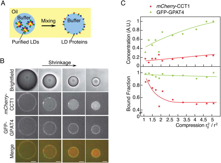
Figure 4. CCT1, but Not GPAT4, Falls Off a Shrinking Oil-Water Interface In Vitro.
(A) Schematic of the in vitro system. LDs in buffer are mixed with TG oil to generate a water-in-oil emulsion. LD proteins then bind to the resulting oil-water interface.
(B,C) During shrinkage of drops in vitro, CCT1 falls off the oil-water interface, whereas GPAT4 remains bound. (B) Representative images are shown. Scale bar, 10 μm. (C) Surface mean concentration and mean surface-bound fraction for mCherry-CCT1 and GFP-GPAT4 are reported. Lines represent trends. A.U. = arbitrary units. See also Figure S2.

