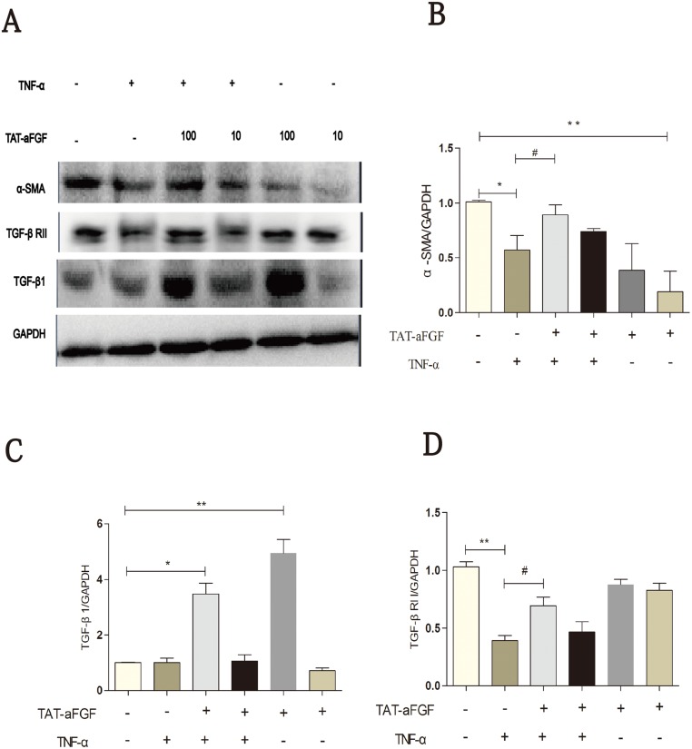
Fig 6. Western blot analysis of α-SMA, TGF-β1 and TGF-βRII in human dermal fibroblasts.
A: Fibroblasts grown in a monolayer were serum-starved for 24 h before the stimulation of TNF-α (5 ng/ml) or TNF-α plus TAT-aFGF (10 ng/ml or 100 ng/ml). Total protein was extracted at the indicated time points. Western blotting was performed for α-SMA, TGF-β1, and TGF-βRII. B, C, D: α-SMA, TGF-β1, and TGF-βRII expression was normalized to GAPDH. Data were obtained from 3independent experiments. * p < 0.05, ** p < 0.01.

