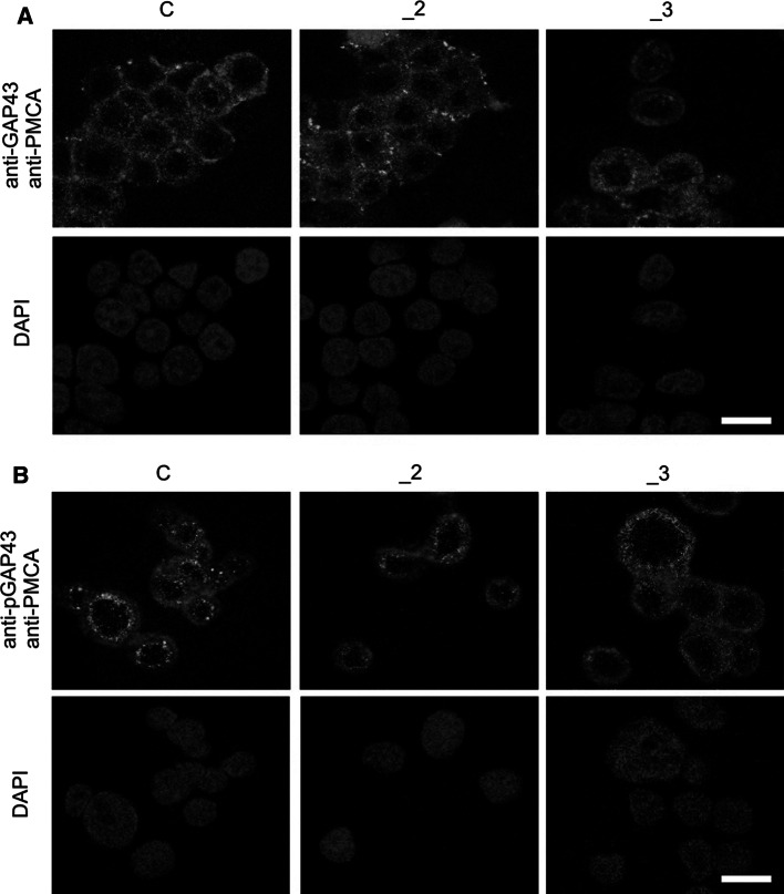
Fig. 3.
GAP43 and PMCA in PMCAs-deficient lines. Representative confocal fluorescent images illustrating a GAP43 and PMCA or b pGAP43 and PMCA co-staining in differentiated PC12 lines. GAP43 or pGAP43 (green) was labeled using protein-specific primary antibodies and anti-rabbit secondary antibodies conjugated with AlexaFluor 488. PMCA (red) was labeled with primary 5F10 antibodies recognizing all isoforms and anti-mouse secondary antibodies conjugated with AlexaFluor 594. Nuclei (blue) were stained with DAPI. Scale bar 20 μm. C control line, _2 PMCA2-deficient line, _3 PMCA3-deficient line. (Color figure online)

