
Figure 1.
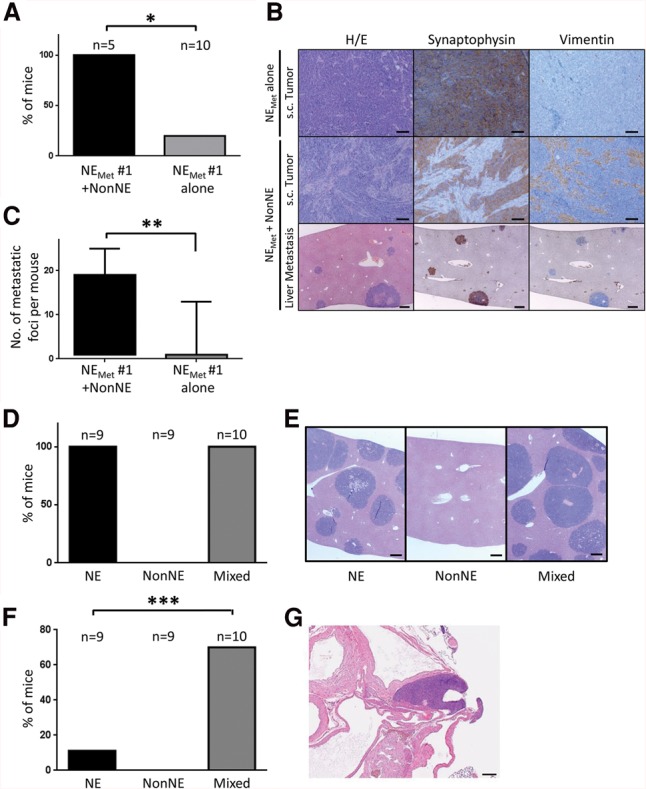
Contribution of NonNE cells to metastasis of NE cells in graft experiments. (A–C) To determine the autonomous metastatic potential of NE cells from metastases, NE cells were established from liver metastases (NEMET cells). NEMET cells were injected subcutaneously into the flank of Balb/c nude mice either as a pure NE cell population or mixed with NonNE cells. (A) The occurrence of metastasis is expressed as the percentage of the number of mice with liver metastases/number of mice in that group. (B) Photomicrographs showing the morphology (H&E staining) and expression of Synaptophysin and Vimentin of transplanted tumors obtained by subcutaneous injection of NEMET cells alone (top panels) and NEMET + NonNE cells (middle panels). The bottom panels show multiple metastatic tumor nodules in the livers of mice injected subcutaneously with NEMET + NonNE cells. Bars: top, middle, 50 µm; bottom, 200 µm. (C) The number of metastatic foci per mouse from each group of mice. Error bars indicate standard deviation (SD). (*) P < 0.005; (**) P < 0.05. (D,E) The supportive role of NonNE cells in metastasis. (D) NE cells and/or NonNE cells were intravenously injected, and liver metastases were evaluated from three independent experiments. (E) Representative micrographs of H&E-stained liver sections. Bars, 200 µm. (F,G) NonNE cells strongly enhance the formation of mediastinal tumors of NE cells. (F) The number of mice showing the mediastinal tumor formation by intravenous injection of both NE and NonNE cells in comparison with injection of only NE cells. (G) Representative micrographs of H&E-stained mediastinal tumor sections. Error bars indicate SD. Bar, 200 µm. (***) P < 0.02.