
Figure 3.
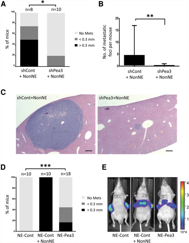
Knockdown of Pea3 in NE cells strongly impairs tumor cell metastasis, and Pea3 overexpression boosts metastasis of NE cells in vivo. (A) The percentage of mice with liver metastases was analyzed by histology. The liver metastases were divided into two sizes: <0.3 mm and >0.3 mm. (*) P < 0.02. (B) The total number of liver metastases was quantified. Statistical significance was determined by Student's t-test. (**) P < 0.01. (C) Representative H&E-stained images of liver sections. Bars, 200 µm. (D,E) A constitutively Pea3- and luciferase-overexpressing NE cell clone was injected subcutaneously into the flanks of Balb/c nude mice. Either control plasmid-overexpressing NE (NE-Cont) cells or mixed NE-Cont and NonNE cells were transplanted as a control group. (D) Statistical significance was determined by Student's t-test. (***) P < 0.01. (E) Representative bioluminescence images for in vivo detection of liver metastasis.