
Figure 4.
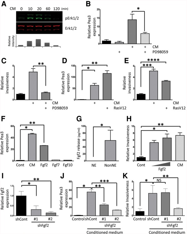
Pea3 expression and invasiveness are induced by fibroblast growth factor (Fgf)/Ras/Mapk pathway activation in NE cells. (A) Erk1/2 activation in NE cells measured after treatment with serum-free conditioned medium from NonNE cells. Samples were immunoblotted with the antibodies against phospho-Erk1/2 (pErk1/2) and total Erk (Erk1/2). The relative intensity of pErk1/2 was normalized to total Erk1/2 using Odyssey software (LI-COR) and is plotted as the fold increase of Erk1/2 phosphorylation as compared with unstimulated NE cells. A similar result was obtained in two independent experiments. (B,C) The effect of MEK1 inhibitor PD98059 on Pea3 expression and invasion of NE cells. (B) NE cells were treated with 50 µM PD98059 and/or conditioned medium from NonNE cells for 12 h, and Pea3 expression was determined by qPCR analysis. (C) NE cells were assayed for their ability to invade Matrigel in the presence of 50 µM PD98059 and conditioned medium from NonNE cells. Data are representative of three independent experiments. (*) P < 0.02; (**) P < 0.02. (D,E) Constitutive Ras activation induces Pea3 expression and invasiveness of NE cells. (D) Lentivirus-mediated overexpression of RasV12 in NE cells induced expression of Pea3 by qPCR in the absence of conditioned medium. (E) Matrigel invasion of constitutive RasV12-expressing NE cells in the absence of conditioned medium treatment. Data are representative of three independent experiments. (*) P < 0.005; (**) P < 0.005; (***) P < 0.01; (****) P < 0.01. (F) Fgf2 induces the expression of Pea3 in NE cells. qRT–PCR was performed to detect the amount of induced Pea3 mRNA in NE cells after treatment with Fgf2, Fgf7, or Fgf10 for 12 h. Data are representative of three independent experiments. (*) P < 0.001; (**) P < 0.0001. (G) Levels of mouse Fgf2 were measured using ELISAs in conditioned medium harvested from NE and NonNE cell clones. Data represent mean ± SEM. Data are representative of three independent experiments. (*) P < 0.01. (H) Effect of Fgf2 (low amount, 1 ng/mL; high amount, 10 ng/mL) on invasion of NE cells in Matrigel. Conditioned medium from NonNE cells was used as a positive control. Data are representative of three independent experiments. (*) P < 0.005; (**) P < 0.001. (I) Inhibition of Fgf2 expression by two distinct shRNA lentiviral constructs in NonNE cells. Data are representative of three independent experiments. (*) P < 0.05; (**) P < 0.005. (J) Conditioned medium from Fgf2 knockdown NonNE cells were used to treat NE cells for 12 h, and Pea3 mRNA expression was measured by qPCR. Data are representative of three independent experiments. (*) P < 0.0001; (**) P < 0.001; (***) P < 0.0001. (K) Quantification of relative invasion of NE cells achieved by conditioned medium from Fgf2 knockdown NonNE cells. Data are representative of two independent experiments. (NS) Not significant. (*) P < 0.005; (**) P < 0.01.