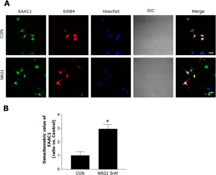
FIGURE 4.
Colocalization of EAAC1 and ErbB4 in primary cortical neurons. A, double immuno-labeling studies on ErbB4 and EAAC1 in primary cortical neurons. Primary cortical neurons were treated with 5 nm NRG1 for 30 min, fixed, and stained with anti-ErbB4 and anti-EAAC1 antibodies that were visualized with FITC and Cy3-coupled secondary antibodies, respectively. Scale bar, 20 μm. B, bar graph summarizing the data from neurons with EAAC1 fluorescence. n = 5. *, p < 0.05.

