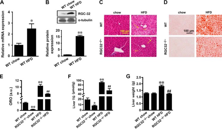
FIGURE 1.
RGC-32 deficiency prevented HFD-induced hepatic steatosis. A and B, RGC-32 mRNA (A) and protein (B) expression in liver tissues of WT mice fed with normal chow or a 12-week HFD (n = 6). C and D, representative images of H&E- (C) and Oil Red O-stained (D) sections of livers from WT and RGC32−/− mice fed normal chow and an HFD as indicated. Microsteatosis and macrosteatosis are indicated by short and long arrows, respectively. E, quantification of Oil Red O staining. ORO, Oil Red O; a.u., arbitrary units. F and G, liver triglyceride (TG) content (F) and liver weights (G) of WT and RGC32−/− mice fed normal chow and an HFD (n = 6). *, p < 0.05 and **, p < 0.01 compared with WT chow groups; ##, p < 0.01 compared with WT HFD groups.

